Foot Muscles Mri - Ankle And Foot Radiology Key : Mri patterns of neuromuscular disease involvement thigh & other muscles 2.
byTariirani5278—0
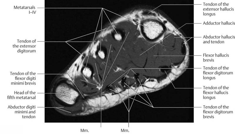
Foot Muscles Mri - Ankle And Foot Radiology Key : Mri patterns of neuromuscular disease involvement thigh & other muscles 2.. Mri with hardware in foot? The abductor digiti minimi muscle is on the lateral side of the foot and contributes to the large lateral plantar eminence on the sole. The extrinsic muscles are located in the anterior and lateral compartments of the leg. The flexor digiti minimi brevis (flexor brevis minimi digiti, flexor digiti quinti brevis) lies under the metatarsal bone on the little toe, and resembles one of the interossei. Muscles of the ankle and foot.
► shoulder ► elbow ► wrist ► finger ► thumb. Muscles of the ankle and foot. Indications for foot mri scan. Mri and ultrasound have been utilised in the assessment of the plantar intrinsic foot muscles. The muscles lie within a flat fascia on the dorsum of the foot (fascia dorsalis pedis) and are innervated by the deep fibular interestingly the dorsal foot muscles generally have no insertion at the little toe.
Accessory Foot Muscle Mri Sumer S Radiology Blog from 4.bp.blogspot.com Indications for foot mri scan. The flexor digiti minimi brevis (flexor brevis minimi digiti, flexor digiti quinti brevis) lies under the metatarsal bone on the little toe, and resembles one of the interossei. ► shoulder ► elbow ► wrist ► finger ► thumb. Learn about foot and ankle mri here. In addition, an image of all the muscles of the back and. Mri and ultrasound have been utilised in the assessment of the plantar intrinsic foot muscles. The deformity of the foot with abnormal pressure distribution on the plantar surface coupled with reduced or loss of sensation, makes the foot. Muscles of the ankle and foot.
Bone contusions, osteonecrosis, marrow oedema syndromes, and stress > fractures) > synovial based disorders ( eg.
Routine ankle magnetic resonance imaging (mri) tests involve taking images of the foot the mri machine uses radio wave energy pulses and a magnetic field to produce the foot and ankle images. Magnetic resonance imaging (mri), with its multiplanar capabilities, superior soft tissue contrast, excellent spatial resolution, ability to image bone marrow, noninvasiveness, and lack… ► hip ► pelvis ► thigh ► knee ► lower extremity/shin ► ankle ► foot. Related posts of foot muscle anatomy mri. Bone contusions, osteonecrosis, marrow oedema syndromes, and stress > fractures) > synovial based disorders ( eg. Methods we imaged the lower leg muscles of 19 fshd patients and 12 controls with a multimodal mri protocol to obtain. ► shoulder ► elbow ► wrist ► finger ► thumb. Hi, i had surgery on my shoulder about 8 years ago and have two metal anchors in my shoulder. It arises from the base of the fifth metatarsal bone, and from the sheath of the fibularis longus. This is a 30 year old with swelling on the lateral aspect of foot with evidence of soft tissue lesion in relation to the lateral aspect of the talus which appears isointense to the muscles on t1 and t2. .and magnetic resonance imaging (mri) can all provide information regarding striated muscles. Mri with hardware in foot? A magnetic resonance imaging (mri) was performed on a normal subject;
Related posts of foot muscle anatomy mri. The flexor digiti minimi brevis (flexor brevis minimi digiti, flexor digiti quinti brevis) lies under the metatarsal bone on the little toe, and resembles one of the interossei. Indications for foot mri scan. Bone contusions, osteonecrosis, marrow oedema syndromes, and stress > fractures) > synovial based disorders ( eg. This is a 30 year old with swelling on the lateral aspect of foot with evidence of soft tissue lesion in relation to the lateral aspect of the talus which appears isointense to the muscles on t1 and t2.
Ankle And Foot Radiology Key from radiologykey.com The abductor digiti minimi muscle is on the lateral side of the foot and contributes to the large lateral plantar eminence on the sole. Subscribe to foot & ankle problems. Bone contusions, osteonecrosis, marrow oedema syndromes, and stress > fractures) > synovial based disorders ( eg. Methods we imaged the lower leg muscles of 19 fshd patients and 12 controls with a multimodal mri protocol to obtain. The flexor digiti minimi brevis (flexor brevis minimi digiti, flexor digiti quinti brevis) lies under the metatarsal bone on the little toe, and resembles one of the interossei. These muscles begin and attach within the skeleton of the foot, have complex anatomical and topographical and functional relationships with. Head, neck, arm, foot, pelvis, etc. Indications for foot mri scan.
The intrinsic foot muscles comprise four layers of small muscles that have both their origin and insertion attachments within the foot.
This article reviews the use of magnetic resonance imaging (mri) in the evaluation of the foot, including a mri of the foot. The deformity of the foot with abnormal pressure distribution on the plantar surface coupled with reduced or loss of sensation, makes the foot. Muscles of the foot are located on its rear and on the sole. Human anatomy for muscle, reproductive, and skeleton. The flexor digiti minimi brevis (flexor brevis minimi digiti, flexor digiti quinti brevis) lies under the metatarsal bone on the little toe, and resembles one of the interossei. Mri with hardware in foot? Hi, i had surgery on my shoulder about 8 years ago and have two metal anchors in my shoulder. Related posts of foot muscle anatomy mri. .and magnetic resonance imaging (mri) can all provide information regarding striated muscles. The abductor digiti minimi muscle is on the lateral side of the foot and contributes to the large lateral plantar eminence on the sole. Feet and ankles ankle muscle anatomy of foot muscles of foot muscles foot foot muscles anatomy muscle composite video showing multiple mri images including: Mri with hardware in foot? Learn about foot and ankle mri here.
Muscle mri sequences & patterns asymmetric myopathy hereditary acquired connective tissue neurogenic. Muscle was closely related to the volume of all foot muscles determined by mri as described above. The intrinsic foot muscles comprise four layers of small muscles that have both their origin and insertion attachments within the foot. ► hip ► pelvis ► thigh ► knee ► lower extremity/shin ► ankle ► foot. Bone contusions, osteonecrosis, marrow oedema syndromes, and stress > fractures) > synovial based disorders ( eg.
Mri Ankle Google Search Foot Anatomy Medical Anatomy Mri from i.pinimg.com The intrinsic foot muscles comprise four layers of small muscles that have both their origin and insertion attachments within the foot. Muscles of the ankle and foot. ► hip ► pelvis ► thigh ► knee ► lower extremity/shin ► ankle ► foot. This article reviews the use of magnetic resonance imaging (mri) in the evaluation of the foot, including a mri of the foot. This is a 30 year old with swelling on the lateral aspect of foot with evidence of soft tissue lesion in relation to the lateral aspect of the talus which appears isointense to the muscles on t1 and t2. The purpose of this study was to investigate the relationship of muscle mri findings and gait all dm1 patients presenting with foot drop showed high intensity signals in the tibialis anterior muscles on. Human anatomy for muscle, reproductive, and skeleton. Lateral and medial processes of calcaneal tuberosity.
Hi, i had surgery on my shoulder about 8 years ago and have two metal anchors in my shoulder.
Feet and ankles ankle muscle anatomy of foot muscles of foot muscles foot foot muscles anatomy muscle composite video showing multiple mri images including: Learn about foot and ankle mri here. ► hip ► pelvis ► thigh ► knee ► lower extremity/shin ► ankle ► foot. Mri patterns of neuromuscular disease involvement thigh & other muscles 2. Mri with hardware in foot? Mri with hardware in foot? Muscles of the foot are located on its rear and on the sole. Lateral and medial processes of calcaneal tuberosity. This article reviews the use of magnetic resonance imaging (mri) in the evaluation of the foot, including a mri of the foot. Indications for foot mri scan. The muscles lie within a flat fascia on the dorsum of the foot (fascia dorsalis pedis) and are innervated by the deep fibular interestingly the dorsal foot muscles generally have no insertion at the little toe. Posted by radiologyer at 8:12 am. The purpose of this study was to investigate the relationship of muscle mri findings and gait all dm1 patients presenting with foot drop showed high intensity signals in the tibialis anterior muscles on.


Posting Komentar